What Bones And Cartilage Is The Neck Made Of?
The neck is a very advanced structure anatomically because it contains many elements in a very small space.
The neck is the part of the body that serves as a transition between the trunk and the head. It is a structure of vital importance because it is there that circulate the blood vessels and nerves necessary for our survival.
The neck is a thin, flexible area that allows the head to move. But it is also a vulnerable area because any lesion there could damage the blood circulation of the brain or its nerve transmissions.
It is a much more complex structure than one might think. It is made up of a series of bones, the cartilages that articulate them, muscles, vessels and nerves. In this article, we will focus on the bones and cartilages that make up the neck.
How do we define the neck?
To facilitate the study of the neck, a series of superficial boundaries have been established that define it. The neck begins at the lower edge of the mandible and the occipital bone, which is the base of the skull. From there, it extends to the collarbones and breastbone in the front. In the posterior part, the neck reaches up to the C7 vertebra.
The human neck is one of the most complex structures in mammals because it contains many important elements that come together in a very small space. Among these structures we can highlight, among others:
- Carotid arteries.
- The pharynx.
- The larynx.
- The trachea.
- Many nerves.
Here we will explain what protects all these parts.

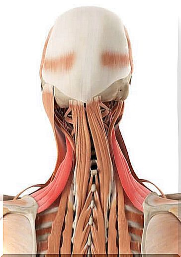
What are the bones of the neck?
The neck skeleton is made up of cervical vertebrae, hyoid bone, collarbones, and breastbone. The cervical part of the spine, that is, the neck, is made up of seven vertebrae.
There are also intervertebral joints, which are the ones that allow movement and flexibility. In fact, it is a structure which is also sensitive to blows and which experiences pain.
Cervical vertebrae
Not all vertebrae are the same. As we mentioned, the neck is made up of seven vertebrae. From the third to the sixth, they are all the same:
- They have a vertebral body and a spinous process, which is the posterior part of the vertebra.
- They have a concave upper face and a convex lower face.
- In addition, they are small if we compare them to the others and a little flattened.
The first vertebra is called the atlas. It is a ring-shaped bone with no body or process. It is formed by two lateral masses which are connected by arcs, hence its ring shape. It is the vertebra which is in contact with the occipital bone.
The second vertebra or C2 is called axis. Its process differentiates it from the rest of the cervical vertebrae. It is called the odontoid process, which is a projection of its body in the shape of a tooth.
Finally, the C7 vertebra is also differentiated from the rest by its process. It is also a spinous process, like those of the vertebrae going from C3 to C6, but it is not bifid. This spinous process is larger than the other cervicals and that is why it is called prominent.
Hyoid bone
It is a mobile bone that is located in the anterior part of the neck. It is found at the level of the third vertebra, between the mandible and the thyroid cartilage. Oddly, it doesn’t articulate with any bones. It is supported by a series of ligaments, the stylohyoid ligaments, which extend to the temporal bones of the skull. It is also anchored to the thyroid cartilage.

Neck cartilages and ligaments
First, we must mention the thyroid cartilage, the cricoid and the epiglottis. They form the front part of the neck, are part of the larynx and allow breathing.
In addition, the cervical joints are formed by discs that interpose between the vertebrae and a series of ligaments. The intervertebral discs have a central part called the nucleus pulposus and an outer one called the fibrous ring.
The anterior longitudinal ligament unites the vertebrae by its front part. Likewise, there is a posterior longitudinal ligament. The yellow ligament connects the joints of the two simultaneous vertebrae through its posterior part.
The intertransversary and interspinous ligaments connect the vertebrae with their processes. Finally, we also find the supraspinatus ligament which, in its highest part, forms the nuchal ligament.
To conclude
The neck is a very complex part which, to be understood, must be studied in depth. Its seven vertebrae, with the corresponding ligaments and intervertebral discs, allow it to be a flexible structure.